

How Innovative Is The Approach To Replace An Xy Animal Model
The winners of our annual 3Rs prize competition, recognising outstanding and original piece of work in 3Rs research.
2020: Dr Laura Pellegrini

Prize winner: Dr Laura Pellegrini, MRC Laboratory of Molecular Biological science
The authors depict a method for developing cognitive organoids representative of the choroid plexus, the protective barrier between the claret and the cerebrospinal fluid (CSF), similar to the claret-brain barrier. The paper, published inScience in July 2020, describes a lot of firsts inin vitro neurological enquiry, from accurately modelling the choroid plexus bulwark office to their ability to produce and store cerebrospinal-like fluid.
Studying the choroid plexus is particularly challenging every bit the region lies deep in the brain, preventing patient biopsies from being used in inquiry. Rodents and macaques are used to establish the ability of drugs to cross the choroid plexus and into the central nervous system, simply many of these drugs neglect in clinical trials. The choroid plexus has previously been modelledin vitro using organoid engineering science, simply the lack of key features including the bulwark function and cerebrospinal fluid production prevented their uptake to replacein vivo studies.
Introducing the human organoids described in the winning paper as anin vitro screening platform could help to prevent ineffective compounds progressing toin vivo studies. They tin as well exist used to written report the power of pathogens to cross into the fundamental nervous system, including the SARS-CoV-two (COVID-19) virus. In addition, the cerebrospinal-similar fluid produced by the choroid plexus organoids contains biomarkers and proteins similar to homo cerebrospinal fluid, with each organoid yielding up to 20 times as much cerebrospinal-like fluid as a mouse embryo.
Pellegrini 50et al. (2020). Homo CNS barrier-forming organoids with cerebrospinal fluid production.Science 369(6500): eaaz5626. doi:x.1126/science.aaz5626
Highly commended: Dr Jennifer Ashworth, University of Nottingham
The highly commended paper past Dr Jennifer Ashworth and colleagues focuses on replacing Matrigel with not-animal-derived hydrogels. Matrigel is derived from mouse tumours and used in manyin vitrostudies to support cell growth. Information technology is increasingly used in 3D cultures for lab-based models of disease, and approximately one hundred mice can exist required to produce the amount of Matrigel needed by a single research plant every twelvemonth. All the same, its chemic components are non well defined, affecting the reproducibility of studies
The hydrogels described in the highly commended paper, published inMatrix Biology, can be 'tuned' by adding proteins and sugars, altering their stiffness and composition to replicate unlikein vivo environments. The hydrogels have been used to support breast cancer cells, including cell lines and patient-derived samples, which would otherwise have needed to be sustained using large quantities of Matrigel, or past implanting into mice.
Ashworth Jet al.(2020). Peptide gels of fully-defined composition and mechanics for probing cell-cell and prison cell-matrix interactionsin vitro. Matrix Biology 85-86: fifteen-33. doi:x.1016/j.matbio.2019.06.009
2019: Dr Francesca Nunn and Dr Marta Shahbazi (Joint winners)

Prize winner: Dr Francesca Nunn, Moredun Inquiry Institute
Poultry cherry mites are a blood feeding ectoparasite. They are a global problem for the egg manufacture, affecting the welfare of laying hens through irritation and anaemia. Mites are controlled using chemicals, however, repeated utilize has led to resistance and recent research efforts have focused on developing vaccines and novel biopesticides. Assessment of vaccine control methods is initially donein vitro using claret assays before field trials are conducted, with 750 to 800 hens exposed to mites for each vaccine candidate, with an adjuvant control group.In vitro assays can be unreliable, for example, due to high levels of not-specific mite mortality. Equally a consequence, vaccine efficacy measuredin vitro is not always translated into mite population reduction in field trials.
Francesca developed an "on-hen" mite feeding device that improves the screening of vaccine candidates to avoid unnecessary field trials. The device consists of a mesh pouch containing approximately 100 mites that have been starved for 3 weeks. The pouch is fitted to the thigh of the vaccinated hen – the mesh is large enough to let the mites' rima oris parts to access the hen'southward skin just pocket-size plenty to comprise the mites. Four hens are used per vaccine candidate and afterward 3 hours, the mesh is removed and the mites are recovered and maintained in 96-well plates for up to six days to appraise mortality. The device has already been used to provide data that has prevented seven vaccines and vaccine commitment methods from going into field trials. The initial pre-screening using the on-hen device involved 56 hens in total, each exposed to 100 mites for 3 periods of three hours – the field studies would take used almost 5,500 birds exposed to x,000 mites for 100 days. The on-hen device has been used past bookish and commercial laboratories in the United kingdom and internationally. By varying the size of the mesh, the device has the potential to exist used in inquiry on other parasites.
Nunn Fet al. (2019). A novel, high-welfare methodology for evaluating poultry red mite interventionsin vivo.Veterinary Parasitology 267:42-46. doi: x.1016/j.vetpar.2019.01.011
Prize winner: Dr Marta Shahbazi, MRC Laboratory of Molecular Biology
Implantation of an embryo into the uterus is a critical step with a high rate of pregnancies lost at this stage. Studying implantation and other early embryonic events is technically and ethically challenging. The majority of work is carried out in mice, typically genetically modified animals where associated surgery and breeding of large numbers of animals are required. Marta'due south enquiry has shown that it is possible to minimise this utilize with reproducible and novel 3D cultures of mouse embryonic stem cells that reliably mimic development at the time of, and beyond, implantation, avoiding the need for recipient mice for embryo transfer and the subsequent alternative of animals to access early stage embryos.
Marta and colleagues accept previously described anin vitro method to civilization human being embryos across the betoken of implantation, overcoming the technical challenges that take traditionally limited the use of human embryos in enquiry. The winning paper builds on this by reporting comparative functional experiments using mouse and man embryonic stem prison cell 3D cultures that have identified key factors involved in the remodelling of the embryo at implantation to form the amniotic cavity. This has revealed a previously unknown link between prison cell authority and tissue shape, with a loss of stalk cell "naïve pluripotency" (that is the ability to become any cell type in the organism) triggering the germination of the cavity and developmental progression of the embryo. The use of the 3D cultures for these studies replaced the use of 500 mice and chiefly past demonstrating that they can be used to respond fundamental biological questions, the research has led to multiple groups worldwide adopting the cultures, further reducing the use of animals.
Shahbazi MNet al. (2017). Pluripotent country transitions coordinate morphogenesis in mouse and human embryos.Nature 552:239-243. doi: ten.1038/nature24675
2018: Dr Rickie Patani
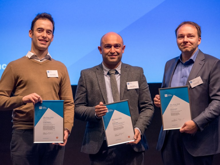
Prize winner: Dr Rickie Patani, UCL Queen Square Institute of Neurology and the Francis Crick Institute
The authors used patient-derived stalk cells to study motor neuron disease, also known as amyotrophic lateral sclerosis (ALS). They optimised a protocol for directed differentiation of induced pluripotent stem cells (iPSCs) into highly enriched (> 85%) functional populations of mature spinal cord motor neurons and astrocytes. Functional and transcriptomic analysis showed that the cells reflect key aspects of ALS pathology, including disrupted motor neuron synapse formation and mislocalisation of nuclear proteins leading to motor neuron expiry.
This model provides a tool to improve understand the illness and to screen potential therapeutics, allowing some studies which are currently dependent on mouse models to be carried outin vitro. Dr Patani and colleagues have already transferred the robust iPSC differentiation protocols to collaborating academic centres, enabling three groups in the Uk to shift some of their enquiry from animal models. The cell model has a number of scientific benefits over the mouse models, which chiefly include the ability to reflect the genetic heterogeneity of patients, investigate sporadic forms of the disease (90% of the cases), and allow early molecular events to exist studied.
Hall CEet al. (2017). Progressive Motor Neuron Pathology and the Function of Astrocytes in a Human Stem Prison cell Model of VCP-Related ALS,Cell Reports 19(nine), 1739-1749. doi:x.1016/j.celrep.2017.05.024
Highly Commended: Dr Diogo Mosqueira, Academy of Nottingham
This paper describes a human being model of hypertrophic cardiomyopathy (HCM), generated by using CRISPR/Cas9 to innovate a disease-causing mutation in human pluripotent stem cells-cardiomyocytes (hPSC-CM). The authors produced 11 variants of mutation in a gene encoding the contractile protein beta myosin heavy chain, often afflicted in the disease. The cells were analysed every bit 2D monolayers and as 3D man engineered eye tissues, which partially restate the architecture of cardiac tissue. Diseased cardiomyocytes showed hallmarks of HCM: hypertrophy and abnormalities in metabolism, contractility and calcium handling.
The cell lines are already being disseminated with three groups in Europe to further build conviction in the model and increase its uptake. In basic research and in early drug development, this cell-based method could exist used to supervene upon some animal models to examine the mechanisms of HCM and efficacy of candidate targets to treat HCM. In toxicology, the cells could exist used to examine the potential for a compound to be harmful to people with HCM.
Mosqueira Det al. (2018). CRISPR/Cas9 editing in human pluripotent stem jail cell-cardiomyocytes highlights arrhythmias, hypocontractility, and energy depletion as potential therapeutic targets for hypertrophic cardiomyopathy.European Heart Periodical 39(43), 3879-3892. doi:10.1093/eurheartj/ehy249
Highly Commended: Dr Bernhard Voelkl, Academy of Bern
The authors investigated how performing experiments in multiple laboratories affects the consequence ofin vivo research, compared to experiments performed in single laboratories. They ran simulations based on 440 preclinical studies across 13 different interventions in animal models of stroke, myocardial infarction and breast cancer, comparing the accurateness of event size estimates betwixt single-laboratory and multi-laboratory study designs across a range of grouping sizes and experimental setups.
Single-laboratory studies mostly failed to predict treatment effect accurately, resulting in an nether- or overestimation of animal group sizes. Using more than animals in single-laboratory studies often resulted in even less valid estimates of result size, as this reduced variability between measurements just did not meliorate the accuracy of the measurements. By contrast, multi-laboratory designs increased accuracy by up to 42% without the demand to use more animals. These findings demonstrate that, despite common assumption, inside-study standardisation is a major crusade of poor reproducibility.
Voelkl Bet al. (2018) Reproducibility of preclinical animal inquiry improves with heterogeneity of report samples.PLoS Biology 16(two): e2003693. doi:x.1371/journal.pbio.2003693
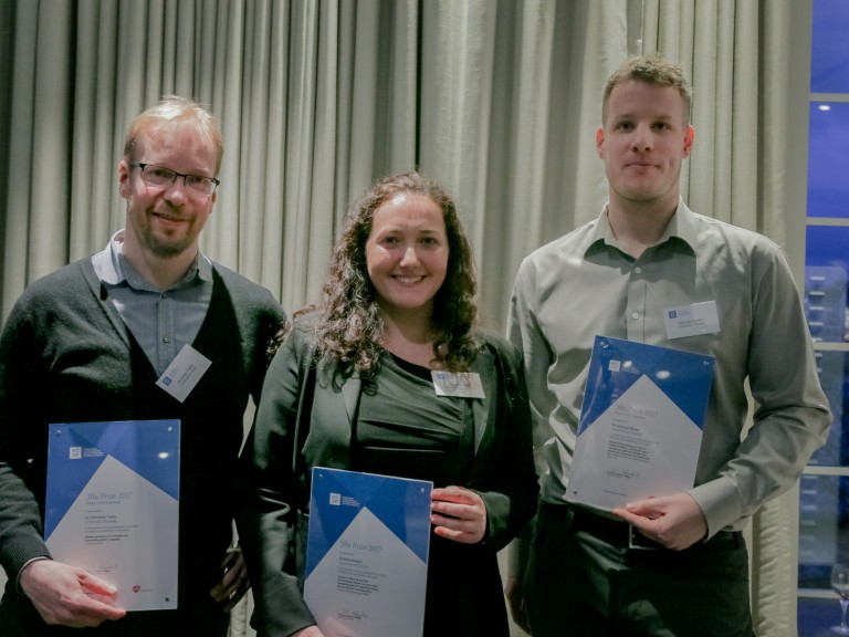
2017: Dr Elisa Passini
Prize winner: Dr Elisa Passini, University of Oxford
The authors developed anin silico model that predicts the risk of drug-induced heart arrhythmias more accurately than animal studies.
They performed an 'in silico drug trial', testing 62 drugs and reference compounds at different concentrations on a control population of 1,213 false human ventricular cells.
Drug-induced changes in heart electrophysiology were measured in a user-friendly software, 'Virtual Assay', developed for this purpose and already used past four pharmaceutical partners. The computer model showed 89% accuracy in predicting the risk of drug-induced heart arrhythmias in humans, in comparison to up to 75% accuracy showed by data obtained from previously conducted animal studies.
Importantly the model likewise has the reward that it represents the electrophysiological variability seen amid patients. It outperforms otherin silico approaches that average experimental information with a one-model-fits-all approach, and do not take inter-field of study variability into account. This consideration of variability in simulations is crucial to capture differences in drug responses on a human population level and holds potential for identifying sub-populations at higher risk.
Passini East, Britton OJ, Lu Hour, Rohrbacher J, Hermans AN, Gallacher DJ, Greig RJH, Bueno-Orovio A and Rodriguez B (2017). Humanin silico drug trials demonstrate higher accurateness than fauna models in predicting clinical pro-arrhythmic cardiotoxicity.Frontiers in Physiology eight: 668. doi:x.3389/fphys.2017.00668
Highly Commended: Dr Christian Tiede, University of Leeds
The paper shows that information technology is possible to generate Affimers, 'culling bounden proteins' to numerous targets, validating the apply of antibody alternative binding reagents in molecular and cellular research studies. They isolated Affimers against 12 different targets and compared them to equivalent antibodies across seven different instance studies. The researchers showed that Affimers, because of their specificity and other factors, can exist equal to, or meliorate than, antibodies for various molecular and cell biology applications.
The power to create Affimers in the laboratory without the use of animals means they could potentially replace (animal-made) antibodies in a number of common molecular and cellular applications.
Tiede C, Bedford R, Heseltine SJet al. (2017) Affimer proteins are versatile and renewable affinity reagents.eLife 2017; six: e24903. doi:10.7554/eLife.24903
Highly Commended: Dr Michael Walker, University of Guelph
The authors depict an alternative experimental design and assay method – termed a 'split-plot' design – where animals are housed in mixed-strain groups. Female C57BL/6 (black), DBA/2 (dark-brown) and BALB/c (white) mice were allocated to either enriched or standard treatments and screened for commonly measured and/ or welfare-relevant behavioural, physiological and haematological variables including corticosterone metabolite output and stereotypic behaviours. Results showed that living in mixed-strain trios did not reduce mouse welfare, or cause any alter to strain-dependent or enrichment typical differences, validating information technology as an alternative study design.
The application of a 'carve up-plot' pattern meant fewer animals were required in each handling grouping. The authors estimate, compared to a traditional design, this design can cut fauna numbers required to achieve 80% ability by more than half. Testing multiple strains increases the external validity of the experiment, thus increasing the generalisability of the results to other ecology contexts or populations.
Walker M, Fureix C, Palme R (2016). Mixed-strain housing for female C57BL/6, DBA/2, and BALB/c mice: validating a split-plot pattern that promotes refinement and reduction.BMC Medical Research Methodology 16: 11. doi:10.1186/s12874-016-0113-7
2016: Dr Joanna Makowska
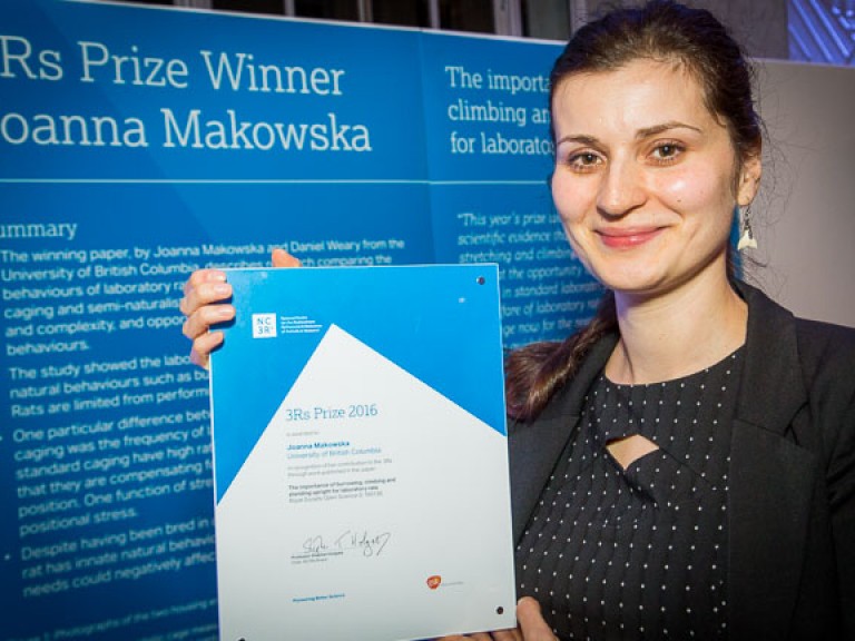
Prize winner: Dr Joanna Makowska, University of British Columbia
The authors provided evidence that natural behaviours such as burrowing and standing upright are of import for the welfare of laboratory rats. They observed the behaviours of rats in 2 dissimilar cage types: standard laboratory caging and big, semi-naturalistic cages with a dedicated expanse for burrowing.
They found that throughout their lives rats burrow readily, fifty-fifty when tunnels are already present, re-organising the tunnels on an almost daily basis. When allowed the space in large cages, rats stand upright multiple times per twenty-four hour period, even until relatively old, and climb often when young. In comparison, rats housed in standard laboratory caging are unable to bear out these behaviours, instead stretching laterally, suggesting that they are compensating for their inability to stretch in the natural upright position.
This work provides a scientific ground for a change in guidelines on laboratory rat housing, including increasing cage meridian and providing burrowing materials. A number of laboratories have already modified their rat housing to conform some of the features described in the newspaper.
Makowska IJ, Weary DM (2016). The importance of burrowing, climbing and continuing upright for laboratory rats.Purple Lodge Open up Science 3: 160136 doi: 10.1098/rsos.160136
2015: Dr Madeline Lancaster and Dr Laura Hall (Joint winners)
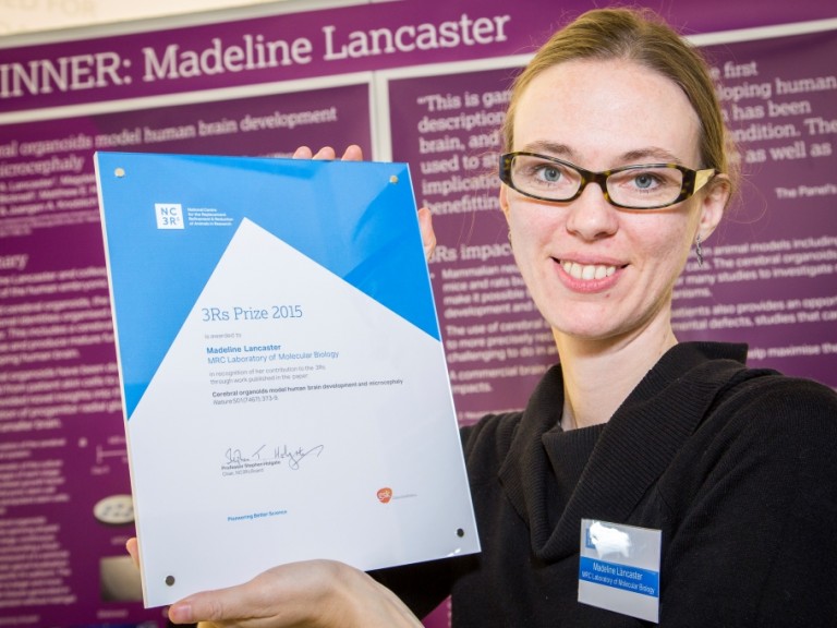
Prize winner: Dr Madeline Lancaster, MRC Laboratory of Molecular Biology
The authors take developed the first 3D model of the homo embryonic brain, using human induced pluripotent stem cells which were able to spontaneously self-organise into a structure that resembles the human brain with discrete, interdependent regions. This was accomplished using adapted growth weather, with specialised matrix support and improved access to nutrients through spinning. The paper also describes using pare cells from a patient with microcephaly to create organoids modelling the illness.
Suitability of animal models in studying neural development is express, every bit they do non recapitulate the anatomical and functional complexity required to study human brain biology and disease. Developing brain organoids from human tissue is a revolutionary footstep towards reducing reliance on animals in studying neurological diseases and potentially in the evolution of new treatments.
- NC3Rs blog: Mini-brains show smashing potential to replace animals in studying neurological disease.
Lancaster Met al. (2013). Cognitive organoids model man brain development and microcephaly.Nature 501(7497): 373-nine doi:x.1038/nature12517
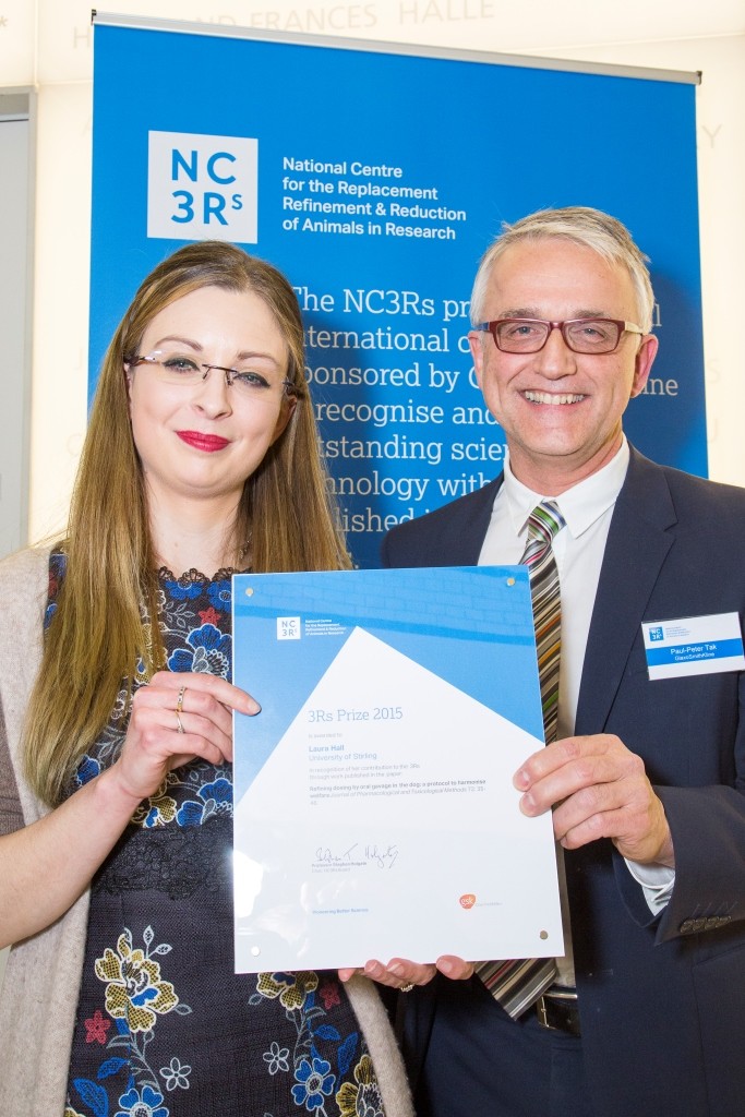
Prize winner: Dr Laura Hall, Academy of Stirling
The study, in collaboration with AstraZeneca, improves the technique of oral dosing in dogs. Following a framework of objective welfare assessments developed by Dr Hall, the authors demonstrate that a modified, refined protocol can minimise stress in dogs compared to the standard approach.
Most laboratory dogs in the Uk are used for condom testing, and oral dosing is 1 of the most common procedures used during these tests. This newspaper shows that using seemingly pocket-size refinements (positive reinforcement grooming with food rewards, a bespeak for dosing, and roofing the dosing tube in palatable paste during grooming) significantly reduces the negative welfare impact of the oral dosing on the dogs. The improved protocol allows researchers to dose dogs more quickly and efficiently, at no further price. Dr Hall has been collaborating with dog facilities beyond the UK to maximise the touch of her work and share all-time practice.
- NC3Rs blog: Improving the welfare of laboratory dogs.
Hall LE, Robinson Due south, Buchanan-Smith HM (2015). Refining dosing by oral gavage in the dog: A protocol to harmonise welfare.Journal of Pharmacological and Toxicological Methods72: 35-46 doi:x.1016/j.vascn.2014.12.007
Highly Commended: Dr Hayley Francies, Wellcome Trust Sanger Institute
The paper describes work on developing and characterising a biobank of colorectal cancer organoids obtained from patients' biopsy tissue. The organoids were shown to accurately reflect the molecular features and diversity seen in the original tumours. More than 80 commonly used or experimental drugs for cancer were tested in the biobank.
Tumour-derived organoids have the potential to replace many studies involving patient-derived xenograft (PDX) mice, including for personalised medicine and drug development. Organoid production is cheaper and has a college success rate than patient neoplasm sample engraftment rates in mice. This technology may fill the gap between cancer genetics and patient trials, complementing and in the long run replacing xenograft based drug studies, and allowing personalised therapy design.
- NC3Rs blog: Cancer organoids: an important new tool in the armoury of cancer biologists.
van de Wetering M, Francies HE, Francis JMet al. (2015). Prospective Derivation of a Living Organoid Biobank of Colorectal Cancer Patients.Jail cell 161(4): 933-45 doi.org/10.1016/j.cell.2015.03.053
2014: Mr Oliver Britton
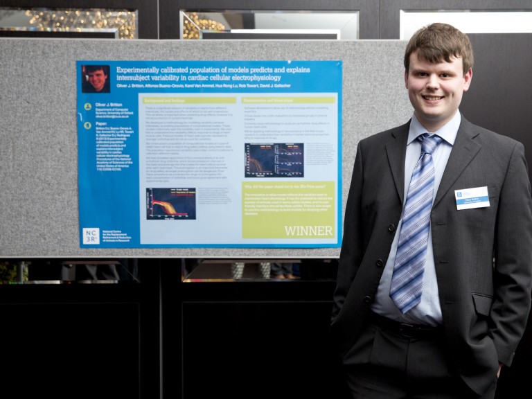
Prize winner: Mr Oliver Britton, University of Oxford
The authors take built a estimator model of cardiac electrophysiology that incorporates natural variability. Normally when using a computer model to examination how a drug might affect the center, the effect of the drug on the center is compared to an average profile of electrophysiology. But this average contour is non really representative of the whole population, where natural variations in heart backdrop occur from person to person. This new approach has the potential to make computer models that are far more than powerful and more predictive of human response, and therefore a more feasible alternative to using animals in inquiry. This is the first time that natural variability has successfully been considered in such a model, and the methodology could be applied to other diseases. The methodology has also been developed into a user-friendly software package chosen Virtual Analysis, which is a major facilitator for manufacture uptake, without the demand for specialist programming and modelling experience. The authors are already planning to use the same methodology to build computer models for agreement pain and diabetes.
Britton O, Bueno-Orovio A, Van Ammel 1000,et al. (2013) Experimentally calibrated population of models predicts and explains intersubject variability in cardiac cellular electrophysiology.Proc. Natl. Acad. Sci. USA 110: E2098–2105 doi:10.1073/pnas.1304382110.
Highly Commended: Dr Olivier Frey, ETH Zurich
Frey'southward paper reports on a novel arroyo to culturing multi-cellular spheroids in vitro. The work is a significant advance in engineering, which brings together the hanging drib method and microfluidics to essentially aggrandize the experimental options for culturing spheroids. Growing cells using a hanging drop approach, means that 3D cell spheroids can be grown without the restriction that may exist imposed by a scaffold or a dish. Microfluidic systems let precise liquid treatment including continuous medium and waste exchange, and also allow examination substances, such every bit candidate drugs, to be run through the culture. This is the first time these two approaches accept been combined, and the outcome holds real promise to eternalize the predictivity of in vitro inquiry. The microfluidic hanging driblet network has already shown that spheroids of cells representing dissimilar organs tin can exist inter-continued in physiological order and are able to communicate with each other via metabolite transfer. This is exciting as this capability is one of the first steps towards creating a multi-organ trunk-on-a-chip model.
Frey O, Misun PM, Fluri DA,et al. (2014) Reconfigurable microfluidic hanging drop network for multi-tissue interaction and assay.Nat. Commun. 5:4250 doi:10.1038/ncomms5250
Highly Commended: Dr Nicola Powles-Glover, AstraZeneca UK
It is a requirement in animal prophylactic testing of new medicines that the blood concentration of the medicine is measured. Historically, big volumes of blood were required to observe the concentration of the medicine. This meant that for rat studies, separate groups of rats were used solely for measuring the concentration while other groups of rats were used to assess the furnishings of the medicine on the fauna. Advances in the way that blood is analysed hateful that, with the right analysis equipment, very modest "microsamples" of claret are now sufficient. Taking a microsample of blood from a rat is a quicker, less stressful process than taking a larger volume. This newspaper provides the show that taking echo microsamples does not adversely affect adult rats and therefore does not interfere with the ability to translate these safety studies. This means that information nigh the drug concentration and its affects can be obtained from the aforementioned animal, allowing a direct link between drug concentration and effect. This is a major scientific improvement. Information technology also ways that far fewer rats are required on these safety studies. As a consequence of this work, whenever the sensitive belittling method is bachelor, AstraZeneca now use microsampling routinely in all rat safety tests.
Powles-Glover Northward, Kirk S, Wilkinson C,et al. (2014) Assessment of Toxicological Effects of Claret Microsampling in the Vehicle Dosed Adult Rat.Regulatory Toxicology and Pharmacology. Apr; 68(3):325-31. doi:10.1016/j.yrtph.2014.01.001
Highly Commended: Drs Brianna Gaskill and Joseph Garner, Purdue Academy and Stanford University
Mice are ordinarily housed at temperatures (xx–24°C) which humans find comfortable for working in the laboratory. Mice get common cold stressed below 30˚C, which can compromise many aspects of physiology and welfare. Withal, the amount of nesting material required to come across a mouse's thermal needs has until now been unknown. In this study the authors plant out where mice wanted to spend their time, based on combinations of temperature and nesting cloth. They found that mice prefer temperatures between 26–29°C, but shift from preferring a warmer temperature to a nest when provided 6-10g of nesting material. These results propose that laboratory mice should be provided with no less than 6g of nesting fabric in order to build fully formed nests, simply 10g or more may be needed to eliminate thermal stress. These results have the potential to positively impact the welfare of millions of laboratory mice all over the globe.
Gaskill B, Gordon CJ, Pajor EA,et al. (2012) Heat or insulation: Behavioral titration of mouse preference for warmth or access to a nest.PLoS ONE vii (3): e32799 doi: 10.1371/periodical.pone.0032799.
2013: Dr Meritxell Huch
Prize winner: Dr Meritxell Huch, Gurdon Institute, University of Cambridge
Dr Meritxell Huch from Cambridge University's Gurdon Institute wins the 2013 3Rs Prize for aNature paper detailing piece of work carried out at the Hubrecht Institute, Kingdom of the netherlands, to develop a civilisation organization that enables adult mouse stalk cells to grow and aggrandize into fully operation three-dimensional liver tissue.
Growing hepatocytes (liver cells) in the laboratory has been attempted past liver biologists for many years, since it would reduce their reliance on using mice to report liver disease and would open upward new opportunities in medical enquiry and drug safety testing. Until at present no laboratory has been successful in deciphering how to isolate and abound these cells.
Liver stem cells are typically found in a dormant land in the liver, only becoming agile post-obit injury to produce new liver cells and bile ducts. Dr Huch and colleagues located the specific blazon of stem cells responsible for this regeneration, which are recognised by a key surface protein (Lgr5+) that they share with similar stalk cells in the intestine, tummy and hair follicles.
By isolating these cells and placing them in a culture medium with the right weather, the researchers were able to abound small liver organoids, which survive and expand for over a year in a laboratory environs. When implanted back into mice with liver disease they continued to grow, ameliorating the illness and extending the survival of the mice.
Having further refined the procedure using cells from rats and dogs, Dr Huch is at present moving onto testing information technology with human being cells, which would not only be more relevant to research into human disease, simply also translate to the development of a patient's own liver tissue for transplantation.
Commenting on the new method'south potential to reduce fauna use in liver inquiry, Dr Huch said:
"Typically a study to investigate one potential drug compound to care for one course of liver affliction would require up to fifty alive animals per experiment, so testing 1000 compounds would need l,000 mice. By using the liver civilization organisation I developed, we tin can test 1000 compounds using cells that come from merely one mouse, resulting in a significant reduction in brute use.
Growing functioning liver cells in culture has been the Holy Grail for liver biologists for many years, so a limitless supply of hepatocytes could have a huge 3Rs affect both on bones research to understand liver illness and for the screening and safety testing of pharmaceuticals.
- NC3Rs weblog: Mini liver enquiry wins 3Rs prize for animal replacement potential.
Huch Met al. (2013).In vitro expansion of unmarried Lgr5 liver stem cells induced past Wnt-driven regeneration.Nature 494: 247252.
Highly Commended: Dr Gyorgy Fejer, Plymouth University
Recognised for the development of a new method to grow macrophage cells for utilise in infectious illness research, which would reduce the employ of mice by many thousands.
- NC3Rs weblog: Immune cells grown in lab could significantly reduce animal utilise in research.
Fejer Yardet al. (2013). Nontransformed, GM-CSF dependent macrophage lines are a unique model to study tissue macrophage functions.PNAS 110(24): E2191-E2198.
Highly Commended: Dr Daniel Adams, University of California San Francisco
Dr Adams has taken inspiration from human orthopaedics to develop a biocompatible, titanium skull implant to reduce infection risk and meliorate welfare in monkeys undergoing cognition studies where encephalon action is monitored directly.
- NC3Rs blog: Taking inspiration from homo orthopaedics to improve research with monkeys.
Adams DLet al. (2011). A watertight acrylic-free titanium recording chamber for electrophysiology in behaving monkeys.J. Neurophysiol 106: 15811590.
2012: Professor Don Ingber
Prize winner: Professor Don Ingber, Wyss Constitute, Harvard University
Professor Ingber's inquiry, published inScience Translational Medicine, describes an innovative 'lung-on-a-chip' microdevice that can accurately replicate conditions in a diseased human lung, offering a viable alternative to using animals in preclinical drug testing.
The microdevice contains hollow channels lined with living human cells, mimicking both the interface between tissues and the unique concrete environment seen in whole living organs. Crystal clear and flexible, it is approximately the size of a USB memory stick.
Applying a vacuum to part of the microdevice allows information technology to 'breathe', recreating the way in which our tissues physically expand and contract during respiration. In testing it was able to successfully replicate the conditions seen in pulmonary oedema (fluid accumulation in the lungs), and predict results of a new drug for this life-threatening condition, which showed benefit in animate being studies.
In addition, the microdevice has allowed the researchers to carry out real-fourth dimension loftier resolution imaging on the cells and make accurate measurements of fluid catamenia and blood jell formation, which are not easily available in an animal model.
Professor Ingber said: "This is precisely the kind of progress that regulatory government agencies, such as the Food and Drug Administration (FDA) in the US, and pharmaceutical companies need to meet in order to seriously consider an culling approach to animal models."
In their paper the researchers depict how the next step is to apply the technology to other homo organs with the goal of i day being able to use it every bit part of an automated system to exam many drugs. While it is not expected to offer an immediate replacement for brute studies, further development and applications of the applied science could let for a more gradual replacement of animal models of man disease.
How the lung-on-a-chip works:
-
Within the microdevice are two parallel, sub-millimeter sized, hollow channels which are separated by a thin, flexible, porous membrane. This membrane is coated with matrix proteins that normally agree cells together in human tissues.
-
One side of this membrane is lined with living human cells isolated from the air sac of a lung, and air is allowed to permeate into the aqueduct to recreate the environs seen in a lung. The other side contains human lung capillary blood cells with a blood-like solution flowing over their surfaces.
-
A vacuum applied to side chambers alongside the channels recreates the way our tissues physically aggrandize and retract when nosotros breathe.
-
Recreating these conditions has been an important step to develop new insights into human lung disease that are difficult to accomplish in with animate being studies, such as the ability to carry out high-resolution imaging on the cells themselves, observing blood clot formation and fluid menstruation.
Huh Det al. (2012). A human disease model of drug toxicity-induced pulmonary edema in a lung-on-a-chip microdevice.Science Translational Medicine 4 (159): 159ra147.
Highly Commended: Professor Susan Barnett, Academy of Glasgow
Scottish-based researcher Professor Susan Barnett was commended for research developing anin vitro model of spinal cord injury using rat embryonic spinal cord cells. This has enabled the laboratory to test the combination of drugs being studied using cells from one creature only, representing a 97% reduction had an established methodology been used. This method is being further developed for testing therapeutics more widely.
Boomkamp SD, Riehle MO, Wood J, Olson MF, Barnett SC (2012). The development of a ratin vitro model of SCI demonstrating the addictive effects of Rho and ROCK inhibitors on neurite outgrowth and myelination.Glia 60 (iii): 441456.
Highly Commended: Professor Gareth Sanger, Queen Mary, Academy of London
London-based Professor Gareth Sanger was commended for research demonstrating the benefit of using man - rather than fauna - gastrointestinal tissues for drug testing, which are obtained equally role of normal surgical procedures.
Broad J, Mukherjee S, Samadi M, Martin JE, Dukes GE, Sanger GJ (2012). Regional- and agonist-dependent facilitation of homo neurogastrointestinal function by motilin receptor agonists.British Periodical of Pharmacology167 (4): 763774.
Highly Commended: Professor Shuichi Takayama, University of Michigan (United states of america)
US researcher Professor Shuichi Takayama was commended for developing a 3D cell culture to test anti-cancer drugs, which proved to be more than representative of clinical responses than standard 2D 'flat' cell cultures, demonstrating the potential for this method to replace and reduce the use of animals in pharmaceutical testing.
- NC3Rs blog: From 2D to 3D, not just a revolution on our TV
Tung YC, Hsiao AY, Allen SG, Torisawa Y, Hoc M, Takayama S (2011). High-throughput 3D spheroid culture and drug testing using a 384 hanging drop array.Analyst136(three): 473478.
2011: Dr Ludovic Vallier
Prize winner: Dr Ludovic Vallier, University of Cambridge
Dr Vallier'southward paper looks at the use of artificial liver cells to model inherited metabolic disorders of the liver has the potential to reduce the number of animals used in this type of research.
The artificial cells, known equally homo induced pluripotent stalk cells (hIPSCs), offer possibilities to regenerate damaged tissues and organs, and information technology is their potential to reduce the number of animals used for screening potential drug treatments that led to Dr Vallier receiving the 3Rs Prize in 2011.
Man liver cells (hepatocytes) cannot exist grown in the laboratory and differences between rodents and humans mean that it is rarely possible to recreate the human affliction completely in mice or rats or to use cultures of rat or mouse liver cells. Dr Vallier'south team took pare cells (dermal fibroblasts) from seven patients with a variety of inherited liver diseases and 3 healthy individuals (the controls). They then reprogrammed cells from the skin samples back into stalk cells. These stalk cells were so used to generate liver cells which mimicked a wide range of liver diseases and to create 'healthy' liver cells from the control group.
Ludovic Vallier's innovative study describes the development and validation of a method to produce cells similar to those in a human liver. Such cells could replace animals for some types of early on drug testing and could also help us to predict adverse clinical reactions. Using these cells for drug testing could be transformative. Ludovic and his colleagues have well illustrated how addressing the 3Rs converges with improving the quality of science.
Rashid TS, Corbineau S, Hannan N, Marciniak SJ, Miranda E, Alexander G, Huang-Doran I, Griffin J, Ahrlund-Richter 50, Skepper J, Semple R, Weber A, Lomas DA, Vallier L (2010). Modelling inherited metabolic disorders of the liver using human induced pluripotent stem cells.The Periodical of Clinical Investigation 120(ix): 3127–3136.
Highly Commended: Dr Anna Williams, MRC Eye for Regenerative Medicine at the University of Edinburgh
Dr Williams' newspaper describes a new jail cell civilization method that dramatically reduces the numbers of mice needed to test potential treatments for nerve cells damanged by multiple scleosis.
In multiple sclerosis (MS), immune cells enter the brain and crusade inflammation and demyelination, where the protective covering of myelin effectually nerves is damaged. The encephalon can repair this impairment past a process called remyelination, but this is non very efficient and frequently fails. Researchers have used rats and mice to try reproduce demyelination and to examination for possible medicines to assist promote remyelination. These experiments utilise large numbers of animals.
Dr Williams discovered that slices of brain taken from very young mice and grown in a dish, retain the 3-dimensional structure and normal cells of the brain and can be used to examination the effectiveness of medicines that might improve remyelination. The researchers likewise automatic the ways in which they measured the remyelination, such that analysis now takes seconds rather than hours.
Zhang H, Jarjour AA, Boyd A, Williams A (2011). Fundamental nervous system remyelination in culture - a tool for multiple sclerosis research.Experimental Neurology230(one): 138–148.
Highly Commended: Dr Stephen Pettit, Wellcome Trust Sanger Institute
Research conducted by Dr Pettitt and colleagues at the Wellcome Trust Sanger Insitute has provided a new manner of generating GM mice which avoids using ii dissimilar strains and therefore reduces the number of animals used.
Scientists use genetically modified (GM) mice to study a range of diseases. But producing GM mice can exist a lengthy process involving large numbers of animals. For technical reasons scientists often use a particular strain of mice for the early parts of the process. However, they then need to breed the mice with another strain to give animals with the desired genetic background for their experiments.
The 'Blackness six' strain of mouse (so-chosen because of its glaze colour) is preferred for many experiments. Nonetheless, for years GM mice could but be made efficiently in a dissimilar strain, '129', from which information technology was easy to derive embryonic stem cells. This meant the GM 129 mice had to be bred with Black half dozen mice for several generations (backcrossing) earlier the mutation could exist analysed on a Black 6 genetic groundwork.
This technique has the potential to reduce the numbers of mice used by hundreds per project. The cells are already in employ in an international project to investigate all twenty,000 mouse genes, and this can at present be done with increased efficiency and fewer animals.
Dr Pettitt'south work consisted of deriving embryonic stem cells the starting point for making a GM mouse directly from Black half dozen mice, thus removing the demand for backcrossing. Importantly, the cells were modified and then that they could exist easily seen by a difference in glaze colour in the mice. This ways that mice with the highest proportion of GM cells tin be selected for convenance ensuring that only those animals likely to transmit the genetic modification to their offspring are used.
Pettitt SJ, Liang Q, Rairdan XY, Moran JL, Prosser HM, Beier DR, Lloyd KC, Bradley A, Skarnes WC (2009). Agouti C57BL/6N embryonic stem cells for mouse genetic resource.Nature Methods vi(vii): 493–496.
2010: Professor Jane Hurst
Professor Jane Hurst's research has shown that a new way of handling laboratory mice can amend their welfare and the quality of the science they are used for.
Laboratory mice are usually picked up past their tails. Professor Hurst's study proves this method of handling causes high levels of anxiety and stress which can influence the outcome of experiments. Past only catching the mice using a plastic tunnel or cupped easily anxiety can exist greatly reduced. This small-scale change tin can be easily applied and has the potential to make a big deviation to the welfare of every mouse used for research.
Hurst, R West (2010). Taming feet in laboratory mice.Nature Methods 7(10): 825–842
2009: Dr Jenny Nichols
Dr Jenny Nichols developed an optimised culture medium for growing mouse embryonic stem (ES) cells and utilised it to derive ES cells from non-obese diabetic (NOD) mice for the first time. This could dramatically reduce the number of animals used to study the genetic basis of blazon 1 diabetes and also has the potential to do the same for mouse models of other diseases.
ES cells are derived from early embryos and can be grown indefinitely in civilization. They are a powerful tool in biomedical research because of their 'pluripotency', which ways they can exist transformed into all the jail cell types in the body, making them widely used inin vitro experiments and to generate disease models in mice.
Previously, agreement which genes play a role in blazon ane diabetes involved convenance NOD mice with strains which had the gene of interest modified. This lengthy procedure required at least ten generations of breeding, involving many hundreds of animals, earlier the mice had a suitable genetic background for conducting the experiment.
The derivation of ES cells from the NOD mouse allows its genes to be directly manipulated to report type 1 diabetes without many generations of backcrossing, dramatically reducing the number of mice required per experiment. The NOD ES cells are now freely available to the research customs, potentially reducing the number of mice used in tpe i diabetes research worldwide.
The new medium, which contains a novel mixture of cell growth factors and inhibitors but no animal products, has also immune the derivation of ES cells from every strain of mouse tested so far with extremely loftier efficiency. Dr Nichols' laboratory has made this applied science available then that it can be applied to other mouse models of disease where deriving ES cells has previously proven impossible.
Nichols J, Jones Chiliad, Phillips J, Newland South, Roode 1000, Mansifeld W, Smith A, Cooke A (2009). Validated germline competent embryonic stem cell lines from non-obese diabetic mice.Nature Medicine 15(seven), 814–818.
2008: Dr Keith Martin and Mr Thomas Johnson
Prize winners: Dr Keith Martin and Mr Thomas Johnson, University of Cambridge
Dr Martin, and his colleague Mr Thomas Johnson, work investigates the potential of stem cells to protect vulnerable nerve cells in the injured retina. Their aim is to develop new treatments for glaucoma, the leading crusade of irreversible blindness worldwide, and other eye diseases. Dr Martin and Mr Johnson pioneered a new method for retinal tissue civilisation that replaces the demand for experiments on live animals.
They have shown that the cultured center tissue remains healthy, maintains its layered architecture, and retains the ability to make new proteins. The tissue also responds to stem prison cell transplantation in a like way to the eyes of living animals.
Besides as replacing the employ of live animals, the new method has brought nigh an viii-fold reduction in the number of animals used, considering eight sections of tissue tin can be obtained from one rat.
Johnson Television receiver, Martin KR (2008). Evolution and characterization of an developed retinal explant organotypic tissue culture organisation as anin vitro intraocular stem jail cell transplantation model.Invest Ophthal Vis Sci 49(8): 3503–3512.
Highly Commended: Mr Charalambos Tymvios, Imperial College London
Mr Tymvious was commended for NC3Rs-funded enquiry on refining a mouse model of pulmonary embolism.
Tymvios C, Jone S, Moore C, Pitchford SC, Page CP, Emerson M (2008). Real-time measurement of non-lethal platelet thromboembolic responses in the anaesthetized mouse. Thrombosis and Haemostasis 99(2): 435–440.
Highly Commended: Dr Jenny Morton, Academy of Cambridge
Dr Morton was highly commended for a paper describing the refinement of tests to measure cognitive deficits in mice used for neurodegenerative disease research.
Morton AJ, Skilings E, Bussey T, Saksida LM (2006). Measuring cerebral deficits in disabled mice using an automated interactive touchscreen organization.Nature Methods 3 (10): 767.
2007: Dr Charlotte Gower
Prize winner: Dr Charlotte Gower, Imperial College London
In her research, Dr Gower worked on schistosomes, the worm-similar parasites that cause bilharzia, a tropical disease affecting an estimated 200 one thousand thousand people worldwide. The affliction can be seriously debilitating, causing long term liver and abdominal damage, and can sometimes be fatal. Dr Gower'southward prize has been awarded for a new application of Deoxyribonucleic acid fingerprinting which replaces the need for using animals in this research expanse and improves the value and accuracy of her results.
Dr Gower has taken advantage of contempo advances in how Dna can exist stored at room temperature and characterised from minute samples in order to collect parasite DNA samples directly from infected people in areas where the disease is owned. Previously, in that location was a need to grow the parasites in the laboratory for study past collecting worm eggs from human being faeces and using them to infect snails and then rodents.
Gower C, Shrivastava J, Lamberton P, Rollinson D, Webster BL, Emery A, Kabatereine North, Webster JP (2007). Evolution and application of an ethically and epidemiologically advantageous assay for the multi-locus microsatellite analysis of Schistosoma mansoni.Parasitology 134: 523–536.
Highly Commended: Dr John Doe, Syngenta
Doe J, Boobis A, Blacker A, Dellarco V, Doerrer Due north, Franklin C, Goodman J, Kronenberg J, Lewis R, McConnell Eastward, Mercier T, Moretto A, Nolan C, Padilla S, Phang W, Solecki R, Tilbury L, van Ravenzwaay B, Wolf D (2006). A tiered approach to systemic toxicity testing for agricultural chemical condom assessment.Critical Reviews in Toxicology 36: 37–68.
2006: Professor Alan Fairlamb and Dr Susan Wyllie
Prize winners: Professor Alan Fairlamb and Dr Susan Wyllie, University of Dundee
In their inquiry, Professor Fairlamb and Dr Wyllie infect hamsters with the parasite Leishmania donovani which causes visceral leishmaniasis. By using a unlike route of infection, the elapsing and severity of the illness in the hamster was reduced without compromising the quality of the scientific upshot.
Professor Fairlamb compared the commonly-used intracardial route of infection with the intraperitoneal road. The results showed that the intraperitoneal route is a simpler, safer and effective method of inoculating the hamsters.
Wyllie Southward, Fairlamb AH (2006). Refinement of techniques for the propagation of Leishmania donovani in hamsters.Acta Tropica 97(three): 364–369.
2005: Dr Siouxsie Wiles
Prize winner: Dr Siouxsie Wiles, Regal College London
In her research, Dr Wiles infects mice with bacteria from the same family unit every bitDue east. coli to study the paths of infection. Traditionally, every mouse has been infected past putting a tube down its throat to deliver the leaner to the stomach - a process chosen gavage.
Dr Wiles tried infecting simply one mouse in this style, then putting it in a cage with uninfected mice and letting nature have its course.
The results showed higher infection rates than the traditional technique. But more importantly, the inquiry was refined so that far fewer animals were subjected to gavage, and the new approach also reduced the full number of animals used by improving the reliability of infection.
- NC3Rs blog: From making bacteria glow to explaining science on the radio
Wiles South, Dougan Chiliad, Frankel G (2005). Emergence of a 'hyperinfectious' bacterial state afterward passage ofCitrobacter rodentium through the host gastrointestinal tract.Cellular Microbiology seven(8): 1163–1172.
Source: https://www.nc3rs.org.uk/3rs-prize-winners
Posted by: martinhambsood.blogspot.com
0 Response to "How Innovative Is The Approach To Replace An Xy Animal Model"
Post a Comment